synaptic transmission and plasticity: spatial and temporal control
The plastic nature of synaptic transmission allows for complex and diverse computations, even with a limited number of neurons in a circuit. Synaptic plasticity occurs on time scales ranging from years to milliseconds. During short-term plasticity, which ranges from milliseconds to minutes, even a single action potential can elicit a several-fold change in a synapse’s response to a second stimulus. The response be weaker, known as short-term depression, or stronger, known as enhancement. The balance of depression and enhancement dictates how all synapses transmit patterns of activity.
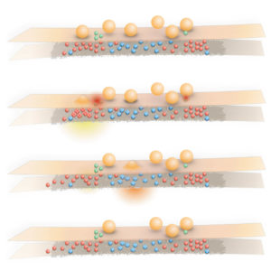
Synaptic transmission is governed by three parameters: 1) the number of synaptic vesicles available for exocytosis, n, 2) the probability that any one of these vesicles will fuse, p, and 3) the postsynaptic current triggered by the neurotransmitter released from one vesicle, q. Modulation in any of these parameters can lead to short-term plasticity. These include the amount of ion flux (i.e. Ca2+, Na+, K+), Ca2+ buffering capacity, sensitivity of vesicles to Ca2+, distance of vesicles to Ca2+ source, and fusion-competence of vesicles. We developed a technique, zap-and-freeze, that couples electrical stimulation with high-pressure freezing to characterize vesicle fusion with millisecond temporal resolution. Using this approach along with protein localization methods, we are currently pursuing the following four projects.
current projects
- spatial and temporal arrangement of release sites within the active zone.
- trans-synaptic organization of release sites with receptors, and their molecular mechanisms.
- molecular mechanisms of short-term plasticity.
- heterogeneity among synapse types
related publications
Ogunmowo, T., Hoffmann, C., Pepper, R., Wang, H., Gowrisankaran, S., Ho, A., Raychaudhuri, S., Cooper, B.H., Milosevic, I., Milovanovic, D., Watanabe, S., (2023) Intersectin and Endophilin condensates prime synaptic vesicles for release site replenishment, DOI: 10.1101/2023.08.22.554276. BioRxiv
Wu, Z., Kusick, G.F., Raychaudhuri, S., Itoh, K., Chapman, E.R.*, Watanabe, S.*, Synaptotagmin 7 supplies docked vesicles for Doc2ɑ-triggered asynchronous neurotransmitter release, BioRxiV. doi:10.1101/2022.04.21.489101. BioRxiV
Kusick, G., Ogunmowo, T., Watanabe, S., (2022) Transient docking of synaptic vesicles: implications and mechanisms. Current Opinion in Neurobiology. 74:102535. link pdf
Vevea, J.D., Kusick, G.F., Courtney, K.C., Chen, E., Watanabe, S., and Chapman, E., Synaptotagmin 7 is targeted to the axonal plasma membrane through γ-secretase processing to promote synaptic vesicle docking in mouse hippocampal neurons, (2021) eLife, 10:e67261, DOI: 10.7554/eLife.67261. link pdf BioRxiv
Li, S., Raychaudhuri, S., Lee, S.A., Wang, J., Kusick, G.F., Prater, C., Syed, S., Falahati, H., Ramos, R., Bartol, T.M., Hosy, E., and Watanabe., S., (2021) Asynchronous release sites are aligned with NMDA receptors at mouse hippocampal synapses. Nature Communications. DOI: 10.1038/s41467-021-21004-x. link pdf BioRxiV
Kusick, G. F. Chin, M., Lippmann, K., Adula, K.P., Davis, M.W., Jorgensen, E.M., and Watanabe, S. (2020) Synaptic vesicles transiently dock to refill release sites. Nature Neuroscience, 23, 1329–1338, DOI: 10.1038/s41593-020-00716-1 link pdf BioRxiV
Return to Research